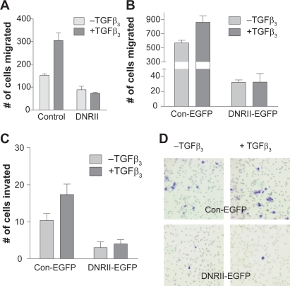
Figure 6.
Effect of DNRII expression on cell migration and invasion. HEC-1-A control and DNRII A) or Control-EGFP and DNRII-EGFP B) cells were seeded in serum free medium in the upper chamber. Culture medium containing 10% FBS with or without 1.0 ng/ml TGFβ3 was added in the lower chamber. To test the invasive potential, HEC-1-A control-EGFP and DNRII-EGFP cells C) were plated in serum free medium onto the upper chamber coated with a layer of Matrigel. After incubation of 20 h for panel A and 24 h for panel B, the cells on the top of the upper chamber membrane were removed. The migrated or invaded cells on the bottom of the membrane were stained with HEMA3 and visualized under a microscope. A representative example is shown in panel D. Quantification was performed by counting the stained cells on each membrane. Values are means ± SME from three chambers.

