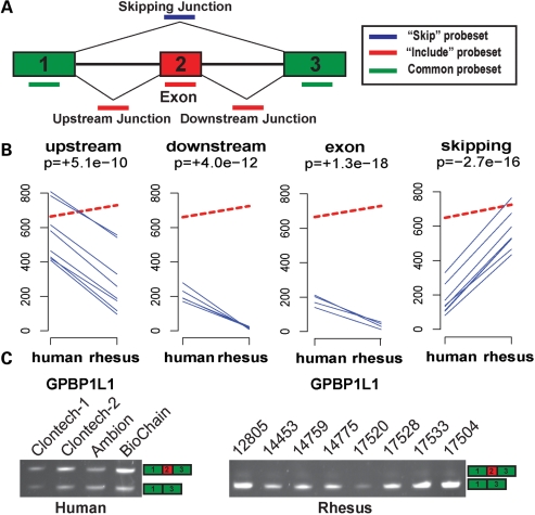
Figure 1.
Detection of a differentially spliced cassette exon between human and rhesus cerebellums by the HJAY array. (A) The HJAY probe design for cassette exons. Each probe set has eight probes. (B) The HJAY array data indicate significantly elevated exon inclusion of GPBP1L1 exon 2 in the human cerebellum compared with the rhesus cerebellum. In each panel, the dashed red line indicates the average gene expression levels of GPBP1L1 in six replicate samples from humans or rhesus macaques. The individual blue lines indicate the average background-corrected intensities of individual probes in a probe set. Only probes that perfectly match orthologous human and rhesus transcripts are shown. MADS+ P-value for differential splicing is shown below the name of the probe set. The ‘+’ and ‘−’ signs indicate the predicted direction of change in exon inclusion levels in the human cerebellum over the rhesus cerebellum. (C) The predicted splicing difference is confirmed by RT–PCR.

