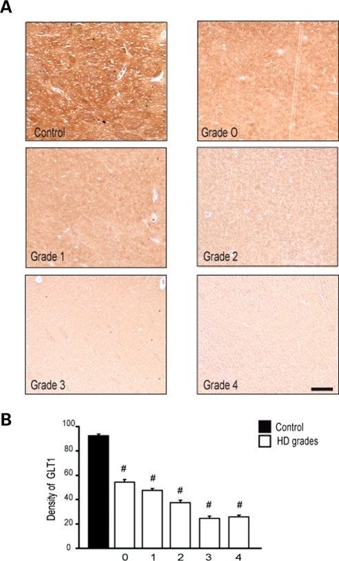
Figure 10.
Immunohistochemical expression of GLT-1 in the caudate nucleus from control and Grade 0–4 HD tissue specimens showed marked reduction of GLT-1 in a grade-dependent manner from HD tissue specimens, in comparison to age-matched normal control specimens (A). Densitometric analysis of GLT-1 immunofluorescence showed significant grade-dependent reductions in GLT-1, with significant differences between grades (B). The magnification bar represents 200 µm.

