Figure 2.
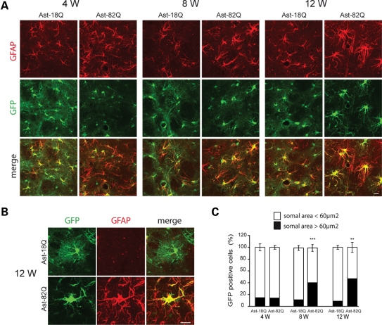
Time course of the morphological changes in astrocytes expressing mHtt. (A) Increase in GFAP immunostaining (red) in astrocytes co-infected with Htt171-82Q or 18Q and GFP (green) at 4- to 12-week post-injection. (B) At 12 weeks, astrocytes expressing mHtt become hypertrophic with larger processes and withdraw most of their finer processes. (C) Quantification of the somal area of GFP-positive astrocytes. At 4, 8 and 12 weeks, the vast majority of the astrocytes expressing Htt171-18Q have a somal area <60 µm2. The proportion changes with time for astrocytes expressing Htt171-82Q, and at 12 weeks, half of the astrocytes have a somal area >60 µm2 (Student's paired t-test versus respective 18Q control, ***P < 0.001; **P < 0.01). Scale bars 20 µm.

