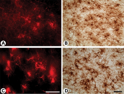
Figure 8.
Immunohistochemistry and immunofluorescence microscopy of the medial area of caudate nucleus (A and B) and putamen (C and D) in a Grade 3 subject. The degree of astroglial dysmorphology was greater in the putamen than in the caudate nucleus, with thickened and tortuous arbors along with increased somal size. The magnification bar in (C) and (D) represents 100 µm.

