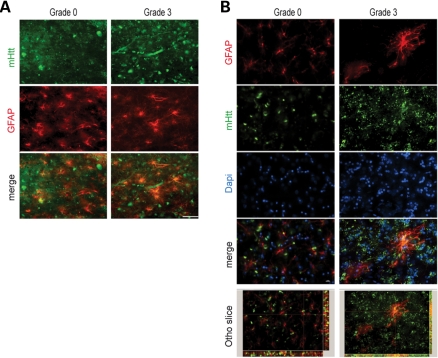
Figure 9.
Combined GFAP and huntingtin immunofluorescence in the medial caudate nucleus in Grade 3 and Grade 0 HD subjects. Two-dimensional analysis showed overlap of each of the antisera (GFAP, red; huntingtin green; and merged figures, yellow) (A). Confocal immunofluorescence of Grade 0 and Grade 3 showed definitive co-localization throughout the tissue specimens (B). The magnification bar represents 20 µm.

