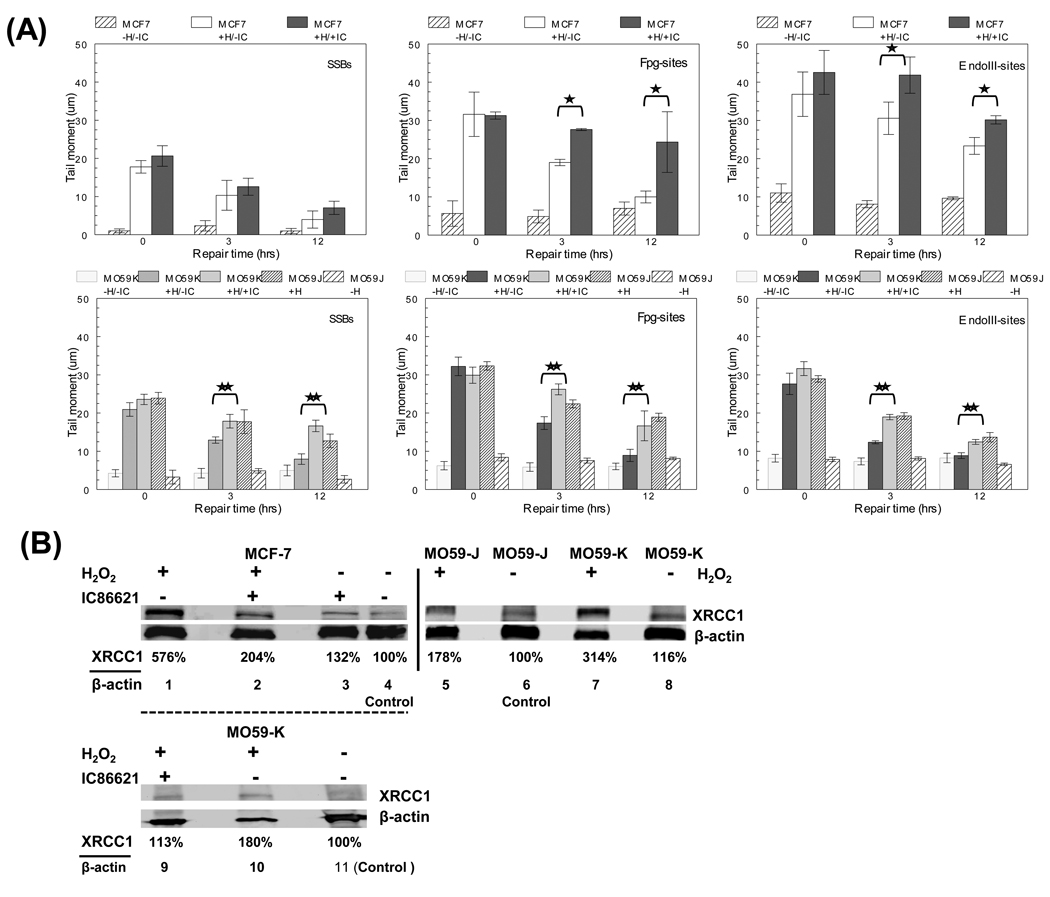
Fig. 3.
Effect of DNA-PKcs inhibition in the processing of single oxidative DNA lesions and base excision repair (BER) pathway (A) Processing of total single DNA lesions in IC86621-treated MCF-7 or MO59-K (+H/+IC: H2O2 and IC86621-treated) and regular MO59J/K cells and controls after exposure for 15 min to 100 µM H2O2 (+H). Detection of SSBs as well as Fpg- and EndoIII-sites using alkaline single cell gel electrophoresis at three post-treatment repair times (‘0’, 3 and 12 hrs). The additional general control no H2O2/IC86621-treated samples has also been included (−H/−IC). Values are averages from two independent experiments. (B) Detection of XRCC1 in IC86621-treated MCF-7 cells 30 min after exposure to 100 µM H2O2 using Western blotting. Forty (40) µg of total protein were loaded/lane. Lanes 1–4, MCF-7 cells; lanes 5–8, M059J/K cells; lanes 9–11 MO59K cells. Lane 4, densitometry control for MCF-7 (DMSO); lanes 6 and 11, MO59J or MO59K (DMSO) with no H2O2 or drug treatment: densitometry controls for MO59J/K. β-actin used as load control. Densitometry values presented as the ratio of XRCC1/actin normalized to control samples. Values averages from two independent experiments. Statistically significant differences between IC86621-treated MCF-7 or MO59-K (MO59K/+IC) and controls (*,** respectively) at p<0.05 are shown.

