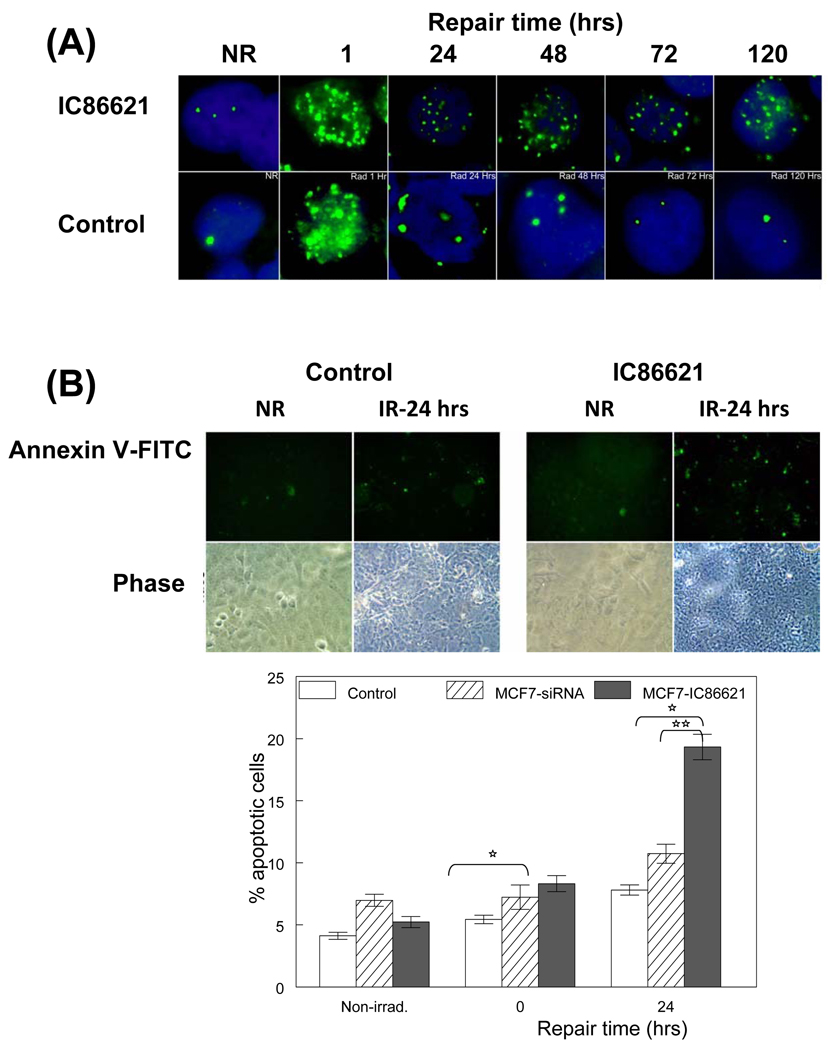
Fig. 4.
Persistence of DNA-PKcs phosphorylated form and apoptosis in MCF-7 cells exposed to γ-rays. (A) phospho-Thr2609 DNA-PKcs foci in IC86621-treated MCF-7 and control cells after exposure to 5 Gy of γ rays. Cells were fixed at different post-irradiation repair times. Both control samples and IC86621-treated were immunostained with rabbit polyclonal to phospho-(Thr2609) DNA-PKcs antibody (green foci). DAPI was used to stain nucleus in all cells. The non-irradiated samples have been included (NR). The results shown here are representative of two independent experiments. (B) Detection of apoptosis using the Annexin V binding assay and fluorescence microscopy at 0 and 24 hrs post-irradiation. Fluorescent and phase pictures for irradiated IC86621-MCF-7 and control cells (IR) as well as non-irradiated (NR) are shown. Annexin V positive cells are green. Quantitation of 200 cells expressed as the percentage of positively stained cells is shown below the representative images. Values are averages from two independent experiments. Values for DNA-PK siRNA cells are taken from Peddi et al. [29] to allow direct comparison. Statistically significant differences between MCF7-IC86621 and controls (*) at p<0.05 are shown.

