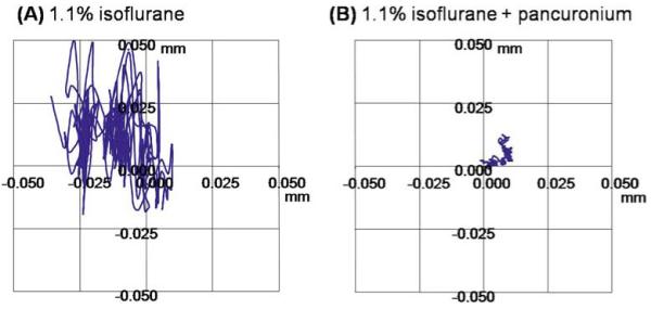
Figure 2.

A, B. Optical imaging of the corneal surface. Optical images were acquired at 7 × 7 μm on an animal under 1.1% isoflurane without (A) and with (B) pancuronium bromide paralytic. The traces show the in-plane displacement of a marker on the corneal surface over 4 min for both experimental conditions. Adapted from figures in Duong et al., J Magn Reson Imaging53 and Zhang et al., Proc Magn Reson Med.83
