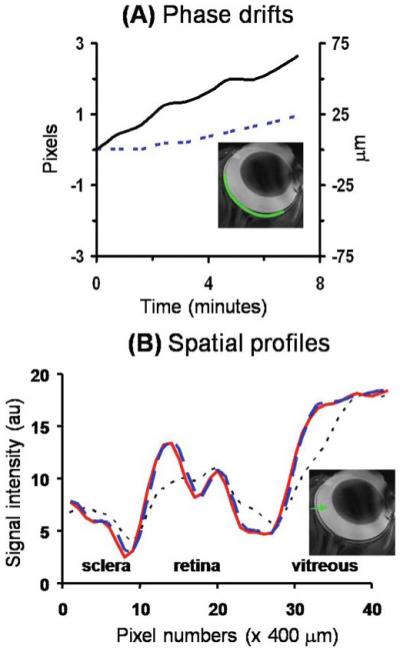
Figure 4.

A, B. In vivo MRI by conventional gradient-echo MRI at 25 × 25 μm. A Temporal-phase evolution in the readout (------) and phase-encode (—) direction of the entire retina obtained from an anesthetized and paralyzed animal using conventional gradient-echo acquisition at 25 × 25 μm. B Image intensity profiles of the in vivo original data (------), after coregistration (– – –), and after phase correction (—) from an in vivo retina. The image profiles were obtained across the retinal thickness from the sclera to the vitreous as shown in the inset. Adapted from figures in Duong et al., J Magn Reson Imaging53 and Zhang et al., Proc Magn Reson Med.83
