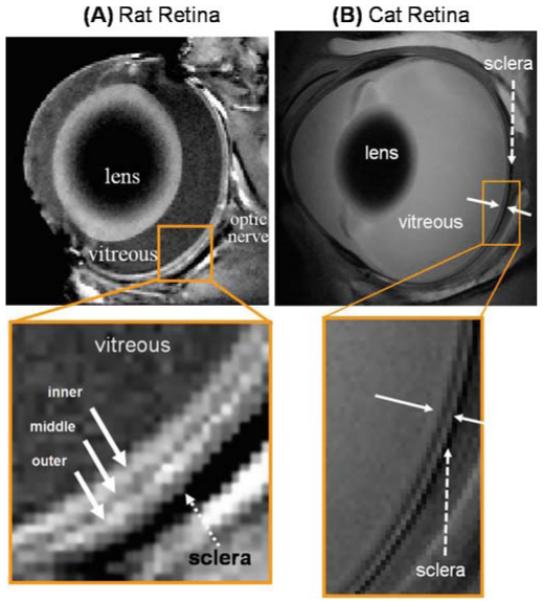
Figure 5.

A, B. Anatomical MRI of rat and cat retina. A Anatomical images from a normal Sprague-Dawley adult rat retina at 60 × 60 × 500 μm. Three distinct layers (solid arrows) of alternating bright, dark and bright bands are evident. The sclera (dashed arrow) is hypointense. Adapted from Fig. 1 of Cheng et al., Proc Natl Acad Sci USA.6 B Cross-sectional T2-weighted (TE = 40 ms) images from a normal cat retina at 100 × 100 × 1500 μm resolution. The solid white arrows indicate the inner and outer strips, respectively. The dashed arrow indicates the hypointense sclera. Adapted from Fig. 3 of Shen et al., J Magn Reson Imaging.48
