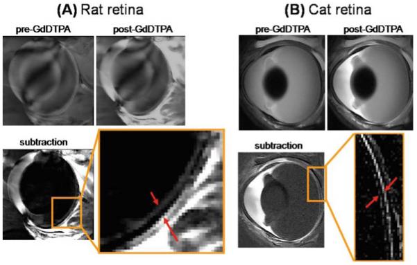
Figure 7A, B.

Gadolinium diethylenetriamine pentaacetic acid (GdDTPA)-enhanced MRI of the rat and cat retina. A Contrast-enhanced images from a normal Sprague-Dawley adult rat retina at 60 × 60 × 500 μm before and after GdDTPA administration and of the subtracted image. The two arrows in the expanded view indicate the inner and outer bands of the retina corresponding to the two vascular layers bounding the retina. Signal enhancement of extraocular tissues supplied by GdDTPA permeable vessels is also present. Adapted from Fig. 2 of Cheng et al., Proc Natl Acad Sci U S A.6 B Contrast-enhanced images from a normal adult cat retina at 100 × 100 × 1500 μm before and after GdDTPA administration and of the subtracted image. The two arrows in the expanded view indicate the inner and outer bands of the retina. Extraocular enhancement was also observed. Adapted from Fig. 6 of Shen et al., J Magn Reson Imaging.48
