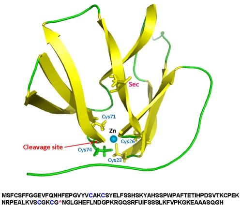
Figure 4. Structural model of mouse MsrB1.
The upper panel shows a structural model of mouse MsrB1. Location of four cysteines in two CxxC motifs and of Sec is indicated. The predicted cleavage site is marked with a red dot and is highlighted by an arrow. Lower panel shows mouse MsrB1 sequence. The two CxxC motifs are highlighted in blue. The predicted cleavage site is also shown as “∧”.

