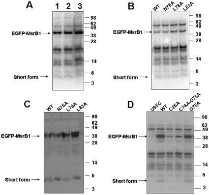
Figure 5. Analysis of the 5 kDa form.
(A) Role of sample preparation in the generation of the 5 kDa form. HEK 293 cells were transfected with pEGFP-MsrB1-F1 for 24 h, then labeled with 75Se for an additional 24 h. Cells were collected and lysed with sample buffer (Lane 3) or Sigma mammalian cell lysis reagent in the presence (lane 2) or absence (lane 1) of protease inhibitors. Protein extracts were resolved on SDS-PAGE, and selenoproteins were visualized with a PhosphorImager. (B-D) Roles of various amino acids in MsrB1 in generating the 5 kDa form. HEK 293 cells were transfected with pEGFP-MsrB1-F1 or constructs containing mutations in one or two amino acids (as shown above lanes) for 24 h, then labeled with 75Se for an additional 24 h. Protein extracts were resolved on SDS-PAGE gels, and selenoproteins were visualized with a PhosphorImager (B, D) or by western blot analysis with MsrB1 antibodies (C). The full-length EGFP-MsrB1 (42 kDa) and the short form (5 kDa) of MsrB1 are shown by arrows.

