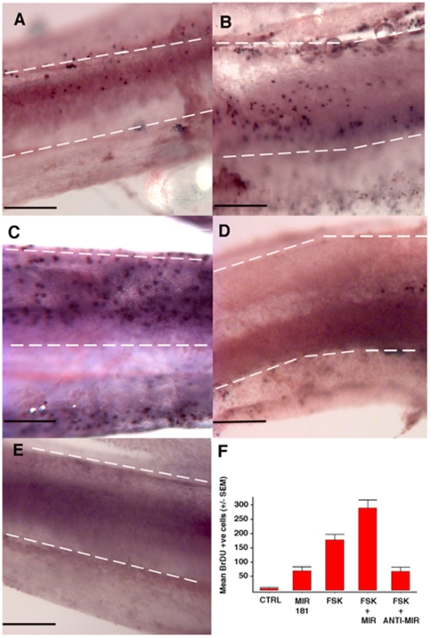
Figure 3. MicroRNA181a overexpression produces proliferation in the chicken basilar papilla.
Basilar papillae from 0-day-old chicks were cultured for 72 hours in the presence of BrdU, the nuclear incorporation of which was detected by antibody labeling. In A–E sensory epithelia are outlined with dashed white lines. The neural edge of each epithelium is at the bottom, the abneural edge is at the top, the distal end is to the right and the proximal end is to the left of the image. BrdU-positive (i.e., recently divided) cells are immunohistochemically labeled and appear as purple dots. Individual cochleas were treated with 100 nM pre-miR181a (A), 100 µM forskolin (FSK) (B), FSK + 100 nM pre-miR181a (C), 100 µM FSK+ 100 nM anti-miR181a (D), or DMSO as a vector control (E). A pro-proliferative effect of miR181a can be appreciated. Further, knocking down endogenous miR181a appears to have a large suppressive effect on forskolin induced proliferation. Transfection was achieved in the first 24 hours of culture using X-tremeGENE siRNA Transfection Reagent in accordance with the manufacturer's instructions (Roche, Indianapolis, IN). The miRNA containing media was removed after 24 hours and replaced with normal medium. The summary of the average number of BrdU positive cells for the control (n = 7), miR181a (n = 8), forskolin (n = 8), forskolin + miR181a (n = 8), and forskolin + anti-miR181a (n = 3) conditions. The following comparisons were all statistically significant: control versus miR181a (p = 0.001), control versus forskolin (p<0.001), miR181a versus forskolin (p<0.001), forskolin versus forskolin plus anti-miR181a (p = 0.008) and forskolin versus forskolin plus miR181a (p = 0.005). Scale bars in panels A–E = 0.2 mm.

