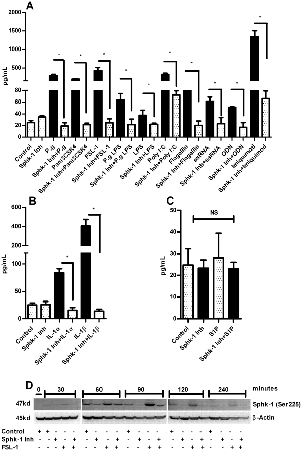
Figure 2. Sphk-1 inhibition down modulates agonists induced HBD-2.
Oral keratinocytes were incubated with or without Sphk-1 inhibitor (2 µM) for 2 h prior to challenging the cells with various TLR agonists namely, heat inactivated P. gingivalis (MOI:100), FSL-1 (1 µg/ml), Pam3CSK4 (0.5 µg/ml), P.gingivalis LPS (1 µg/ml), E. Coli LPS (1 µg/ml), ssRNA (0.1 µg/ml), Poly I:C (5 µg/ml) ODN (0.5 µg/ml), Imiquimod (0.1 µg/ml), Flagellin (0.25 µg/mL) (A); the IL-1α (2.5 ng/ml), IL-1β (2.5 ng/ml) and TNF-α (2.5 ng/ml) (B) and GPCR agonist S1P (100 nM) (C) for 24 h. The supernatant was collected after 24 h and HBD-2 ELISA was performed using Human BD-2 ELISA kit. The Sphk-1 inhibitor ablated HBD-2 induction with the agonists tested. The time course experiment was performed by pretreating the cells with Sphk-1 (2 µg/ml) for 2 h before challenging with FSL-1 (1 µg/ml) for 0, 30, 60, 90, 120 and 240 min. The total protein was collected and subjected to immunoblot with ser225 phospho specific Sphk-1 antibody and β-actin as loading control. We noted increase in the phosphorylation level of Sphk-1 at ser225 as early as 60 min and the level of phosphorylation was down regulated in the presence of Sphk-1 inhibitor (D). Control cells received DMSO unless otherwise stated. Results are mean ± SEM and are representative of three independent experiments. Statistical comparisons are shown by horizontal bars with asterisks above them (* indicates p<0.05 determined by ANOVA and Tukey multiple comparison test).

