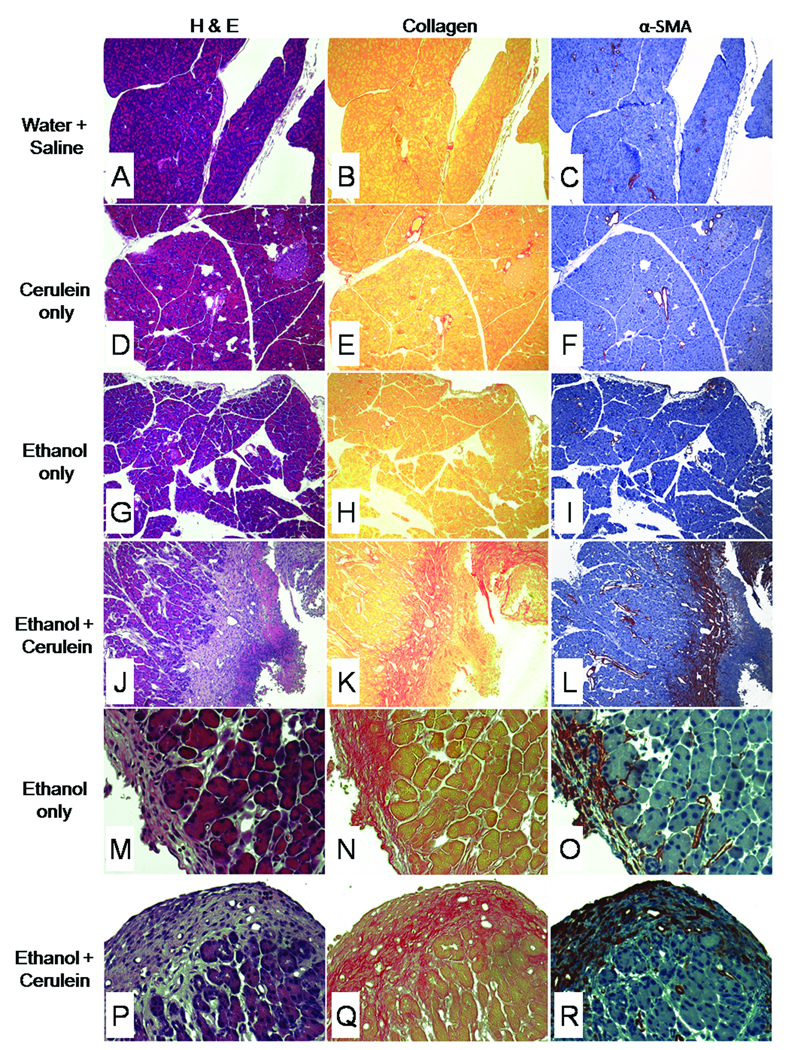
Figure 2. Histological changes in mouse pancreatic tissues after chronic administration of ethanol.
Mice (n= 6 per group) received water/saline (A–C), cerulein alone (D–F), ethanol alone (G–I, M–O) or ethanol plus cerulein (J–L, P–R) for 3 weeks as described in Materials and Methods. Pancreatic tissue sections were subsequently stained with H&E (A,D,G,J,M,P), Sirius red (B,E,H,K,N,Q) or anti- α-SMA (C,F,I,L,O,R). Pancreata from mice that were treated with water/saline (A–C), cerulein alone (D–F) showed no overt pathology, with no evidence of increased collagen deposition or increased α-SMA production. In these mice, collagen and α-SMA were restricted to, respectively, the supporting structure and smooth muscle lining of the vasculature. Mice treated with ethanol alone showed no overt pathology other than minor collagen deposition and αSMA staining in focal areas of peripheral fat tissue (M–O), which was also seen in mice treated with ethanol plus cerulein (P–R). In contrast, pancreata of mice treated with ethanol plus cerulein (J–L) showed massive collagen deposition and a highly correlated and intense amount of α-SMA staining in and around areas of acinar degeneration and necrosis. (x100:A–L, x400:M–R).

