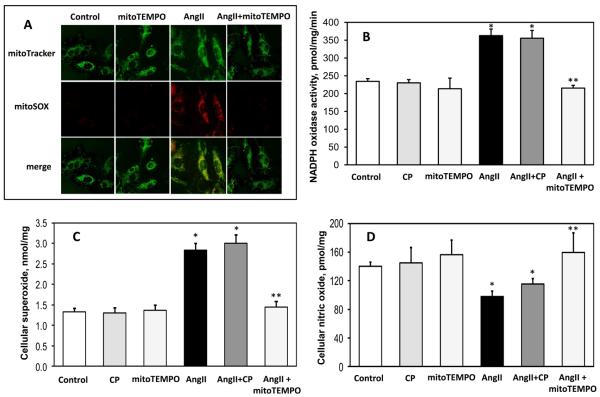
Figure 2.
Effect of mitoTEMPO on mitochondrial O2∸, endothelial O2∸, nitric oxide and NADPH oxidase activity. (A) Mitochondrial O2∸ was measured in control or angiotensin II (Ang II)-stimulated HAEC using fluorescent probe MitoSOX. Mitochondrial localization of MitoSOX signal was confirmed by colocalization with MitoTracker. (B) Activity of NADPH oxidase measured in membrane fractions isolated from unstimulated or angiotensin II (Ang II) stimulated BAEC (4 hours, 200nM) and supplemented for 15 minutes with saline, the mitochondria-impermeable SOD mimetic 3-carboxyproxyl (CP), or the mitochondria-targeted SOD mimetic mitoTEMPO (25 nM). (C) Cellular O2∸ was measured in intact BAEC using DHE and HPLC. (D) Nitric oxide was measured in intact cells after treatment with saline, CP or mitoTEMPO using ESR and the NO spin trap Fe(DETC)2 24. Results are mean±SEM, n=5-8 each. *P<0.05 vs control, ** P <0.05 vs Ang II.

