Fig. 4.
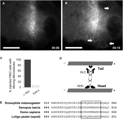
Microtubule sliding occurs in a variety of cell types, and is driven by KHC in Ptk2 cells. (A) Xenopus fibroblast transiently expressing Dendra2-α-tubulin immediately after photoconversion in a narrow bar; (B) the same cell approximately 5 min later. (Scale bars, 20 μm.) Arrows point to fluorescent microtubule segments that moved away from the converted region. (C) Percent of PtK2 cells injected with SUK-4 or a control antibody (mouse anti-myc) possessing moving microtubules as compared with uninjected cells (motility determined in a double-blind survey). (D) Model of kinesin-1–driven microtubule-microtubule sliding. (E) Alignment showing conservation of C-terminal microtubule binding domain (boxed region) identified in Drosophila KHC (19).

