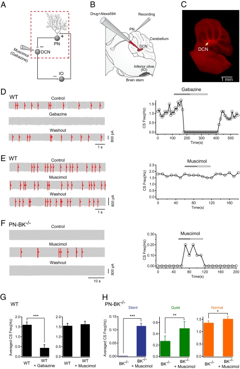
Fig. 4.
Rescue of CS activity in PN-BK−/− mice by increasing inhibition in the DCN. (A) Schematic presentation of the olivo-cerebellar circuit (Left) and the segment under examination (red dotted square) when gabazine or muscimol was locally applied to the DCN. (B) Experimental configuration for cell-attached recordings from PNs and local drug applications to the DCN. The glass pipette for drug application filled with Alexa594 was lowered from cortex into the DCN. (C) Fluorescence image showing the site of local drug application within the DCN (arrow). (D) Representative electrical traces and time-course of the effect of gabazine on CS activity in a WT cell. Gabazine (200 μM) applied into DCN dramatically reduced the frequency of CS. (E) Representative traces and time course showing the absence of effect of muscimol (300 μM) on the CS frequency in a WT mouse. (F) The CS activity was restored in a silent PN-BK−/− cell during application of muscimol into DCN. (G) Summary of D and E (n = 8 cells for gabazine experiments, n = 11 cells for muscimol experiments; paired t tests). (H) Summary of the effect of muscimol on three classes of CS activity in PN-BK−/− mice: silent (n = 7 cells), quiet (n = 7 cells), and normal (n = 5 cells) (paired t tests). *P < 0.05; **P < 0.01; ***P < 0.001. Error bars show SEM.

