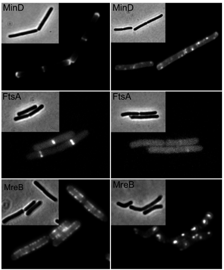
Fig. 1.
Pmf-dependent localization of proteins. Cellular localization of GFP-MinD, YFP-FtsA, and GFP-MreB in B. subtilis cells in the presence (Right) and absence (Left) of the proton ionophore CCCP (100 μM). Strains used: B. subtilis 1981 (GFP-MinD), B. subtilis PG62 (YFP-FtsA), and B. subtilis YK405 (GFP-MreB).

