Fig. 1.
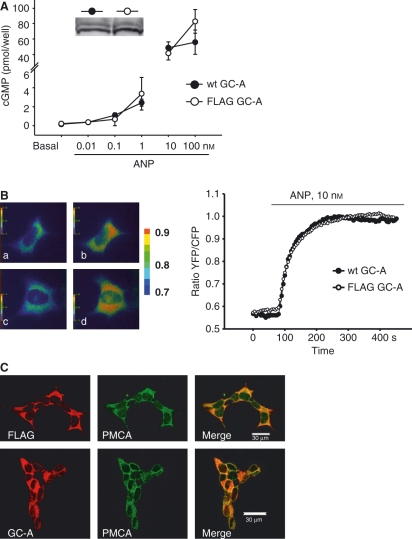
The N-terminal FLAG epitope does not alter the activity and subcellular localization of the GC-A receptor. (A) HEK293 cells expressing either the wild-type (wt) GC-A or FLAG-tagged GC-A receptor were incubated with ANP (10 pm to 100 nm, 10 min). Intracellular cGMP contents were quantified by RIA. Inset in (A): western blot analysis demonstrated similar expression levels of wt and FLAG-tagged GC-A. (B) FRET was used to monitor the kinetics and extent of cGMP formation in single HEK293 cells cotransfected with either wt GC-A or FLAG-tagged GC-A and cGMP indicator (pGES-DE2 [24]). Left: FRET images of two cells prior to and during incubation with ANP: wt GC-A with vehicle (a) and 10 nm ANP (b); FLAG-tagged GC-A with vehicle (c) and ANP (d). Right: representative ratiometric recordings of single-cell FRET signals. (C) Confocal immunofluorescence images of HEK293 cells transfected with wt or FLAG-tagged GC-A demonstrate the colocalization with PMCA.

