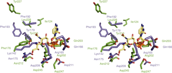
Fig. 4.
Cross-eye stereo-view of the residues of interest (blue) in the active site of the CNS from Neisseria meningitidis containing CDP (yellow) (PDB 1EYR) [12] superimposed with their equivalent residues (green) of the murine CNS containing CMP-Neu5Ac (yellow) (PDB 1QWJ) [18]. All residues are numbered, with the exception of Gln104 in the N. meningitidis CNS equivalent to Gln141 in the murine enzyme, which are shown at the back of the view behind the sialic acid molecule.

