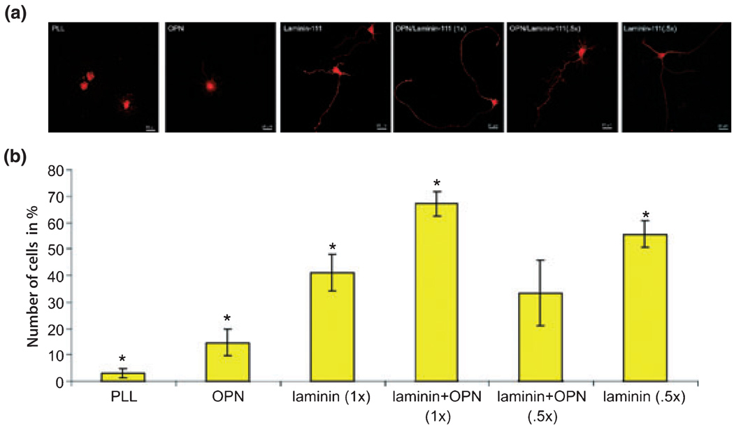
Fig. 2.
RGCs plated on different substrates and characterized respectively by their axon length on these substrates. (a) Confocal images of RGCs plated on poly-l-lysine (PLL), osteopontin (OPN), laminin (1×), OPN/laminin (5×), OPN/laminin (1×), and laminin (0.5×) mixtures. (b) The percentage of RGCs that extended axons in vitro was quantified on the aforementioned substrates (5 wells/condition). RGCs adhered to all substrates; *statistically significant, p < 0.05.

