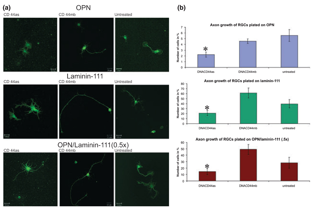
Fig. 4.
CD44 down-regulation decreases RGC axon initiation response. (a) Confocal images of RGCs plated on osteopontin (OPN), laminin-111, or mixture of OPN/laminin-111 (0.5×) treated with a deoxyribozyme against CD44 (as, first column), control deoxyribozyme (mb, second column), or left untreated (third column) for 1 day and immunostained for β-tubulin. (b) Quantification of the percent of RGCs that initiated axons on each substrate with deoxyribozyme or control treatments. The use of percentages were justified by increased linearly (no-intercept model) between the number of axonal cells compared to total number of cells counted. *p < 0.05 by the General Linear Model procedure (SAS Software, Cary, NC, USA), and by subsequent pair-wise tests using Fisher’s LSD-test.

