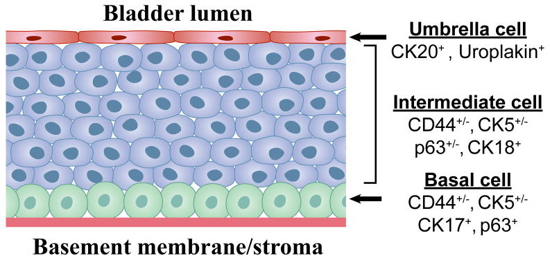
Figure 4. Differentiation-based lineage hierarchy in the normal urothelium.
The normal urothelium is comprised of 3 distinct layers. The surface epithelium consists of a single layer of terminally differentiated urothelial cells (“umbrella cells”) that express uroplakins and cytokeratin-20 (CK20) and have a low proliferative potential. It is conceivable that papillomas arise from transformation of umbrella cells, but this would require that they “de-differentiate” to reacquire self-renewal potential. Below the umbrella cells is a multi-cellular layer of intermediate cells that express CK18 and variable levels of p63, CK5, and CD44. These are most likely “transit-amplifying cells” that possess higher proliferative potential but have also begun to differentiate towards the umbrella cell phenotype. Finally, adjacent to the basement membrane is a layer of basal cells that express high levels of CK5, CD44, p63 and the 67 kD laminin receptor. This layer contains urothelial stem cells, which are tightly regulated by stromal elements (cells and matrix proteins) contained within the “niche”. Recent studies indicate that urothelial cancer stem cells share several molecular markers with the normal basal cell, including CD44, CK5, and the 67 kD laminin receptor and that these markers are upregulated at the tumor-stromal interface in xenografts.

