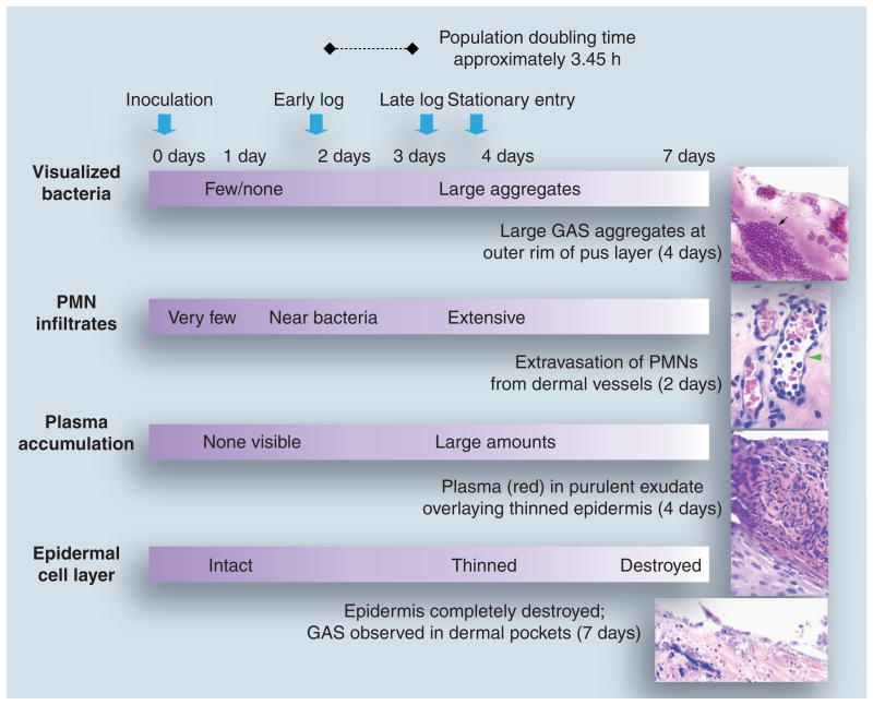
Figure 3. Growth dynamics of group A Streptococcus during skin infection in the humanized mouse.
Pathological alterations throughout the time course of GAS infection of skin grafts in the humanized mouse are described. The time scale (top) designates the day postinoculation of skin with GAS. The growth stage of infecting GAS, determined based on the number of colony-forming units recovered from the skin at different time points, is also described. Panels on the right represent hematoxylin- and eosin-stained tissue sections obtained from GAS-infected skin biopsies.
Black arrow: GAS aggregates; green arrowhead: dermal capillary containing PMNs.
GAS: Group A Streptococcus; PMN: Polymorphonuclear leukocyte.
Tissue sections reproduced with permission © American Society of Microbiology.

