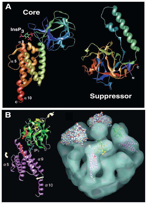
FIG. 4.

Structures of the InsP3R. A: crystal structures of the core InsP3 binding domain (left) and suppressor domain (right). InsP3 present in the core domain structure coordinated in a cleft created by an NH2-terminal β-sheet-rich β-trefoil domain and an α-helical armadillo-repeat domain. Suppressor domain is comprised entirely of a β-trefoil domain (head) with a helical insert (arm). Structures solved in Refs. 52, 54. B: cryoelectron microscopic single-particle reconstruction of InsP3R (right, tilted with respect to the plane of the page with cytoplasmic aspect facing upwards toward viewer with InsP3 binding core domain density fitted into an L-shaped density). For a better fit, various parts of the InsP3 binding core domain were rotated as indicated by the arrows with respect to the crystal structure shown in A. N and C refer to NH2 and COOH termini of each domain in A and B. [From Sato et al. (407), with permission from Elsevier.]
