Fig. 5.
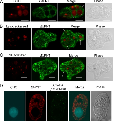
Localization of EhPNT to phagosomes, lysosomes, and endosomes. (A) Association of EhPNT with phagosomes. Amoebae were incubated with CellTracker Orange-loaded CHO cells (red) for 60 min, fixed, and reacted with anti-EhPNT antibody (green). Arrows indicate representative phagocytosed CHO cells associated with EhPNT. (B) Association of EhPNT with lysosomes. Amoebae were labeled with LysoTracker (red) and subjected to an immunofluorescence assay with anti-EhPNT antibody (green). Arrows indicate representative lysosomes associated with EhPNT. (C) Association of EhPNT with the fluid-phase marker. Amoebae were incubated with medium containing RITC-dextran (red) for 1 h. The cells were fixed and reacted with anti-EhPNT antibody (green). Arrows indicate representative endocytosed RITC-dextran associated with EhPNT. (D) Subcellular localization of phagosomes, mitosomes, and EhPNT. The amoebic transformant expressing EhCpn60-HA was incubated with CellTracker Blue-loaded CHO cells (blue) for 60 min, fixed, and reacted with anti-EhPNT (red) and anti-EhCpn60 (green) antibodies. Bar, 10 μm.

