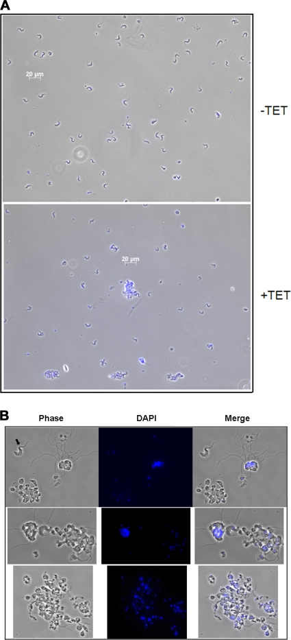
Fig. 7.
Microscopy of bloodstream form cells exhibiting aberrant morphologies upon TbPRMT6 depletion. (A) Morphological changes evident in BF TbPRMT6 RNAi cells. Cells grown in the presence (+TET) or absence (−TET) of tetracycline for 6 days were fixed to slides, stained with DAPI, and analyzed by microscopy. (B) Images of the giant cells with detached flagella evident in BF TbPRMT6 RNAi cells. Note the BF cell with normal morphology (black arrow) for scale reference.

