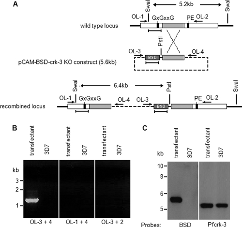
Fig. 4.
Attempt at disrupting the pfcrk-3 gene. (A) Strategy for disruption of the pfcrk-3 gene. The transfection plasmid contains a PCR fragment spanning positions 1277 to 2368 of the 4.2-kb pfcrk-3 coding sequence. This fragment excludes two kinase subdomains essential for activity, labeled GXGXXG (a glycine-rich region required for correct orientation of ATP) and PE (a proline-glutamate motif in which the latter residue is required for the structural stability of the enzyme). Single-crossover homologous recombination results in a pseudodiploid configuration with two truncated copies, each of which lacks one of these essential motifs. Oligonucleotides are indicated by horizontal arrows, restriction sites by vertical lines, and DNA probes by horizontal bars. BSD, blasticidine deaminase cassette. (B) PCR analysis of the disrupted locus. Total DNA isolated from cloned, blasticidin-resistant parasites transfected with pCAM-BSD-crk-3 (transfectant) or from wild-type 3D7 parasites (3D7) was subjected to PCR using the primers indicated (see panel A for their locations). (Left) Primers OL-3 and -4 (diagnostic for the pCAM-BSD-crk-3 episome or concatemeric inserts); (center) primers OL-1 and -4 (diagnostic for 5′ integration); (right) primers OL-3 and -2 (diagnostic for 3′ integration). (C) Southern blot analysis. Total DNA was extracted from cloned blasticidin-resistant parasites transformed with pCAM-BSD-crk-3 (transfectant) and from wild-type 3D7 parasites (3D7) and was digested with PstI and SwaI. (Left) After transfer to a Hybond membrane, the digested DNA was probed with the blasticidin resistance cassette. (Right) The membrane was stripped, and the digested DNA was probed with a pfcrk-3 amplicon located upstream of the fragment used as an insert in the pCAM-BSD-crk-3 plasmid. The sizes of comigrating markers are given to the left. The band corresponding to the linearized episome was detected in the transfected parasites (left panel, left lane). The band corresponding to the intact wild-type locus was detected in both the transfected and the untransfected parasites (right panel, both lanes).

