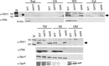
FIG. 6.
PilY1 cellular localization. Cellular fractions of the WT, ΔpilY1 mutant, and ΔpilA mutant were separated by SDS-PAGE. Fractions are indicated as supernatant (Sup), cell-associated (CA), whole-cell (WC), soluble cytoplasmic (Cyt), total membrane (TM), inner membrane (IM), and outer membrane (OM) fractions. The cell-associated fraction refers to proteins weakly associated with the cell surface that can be released by brief vortexing. Western analysis was performed using the following antibodies as indicated: anti-PilY1, anti-SecY, anti-OprF or anti-PilA antibody. SecY (∼50 kDa) served as a control for inner membrane localization, and OprF (∼37 kDa) served as an outer membrane marker. The protein size marker lanes are indicated (M), with sizes in kDa. The arrow indicates the position of the PilY1 protein at ∼110 kDa. The molecular mass of WT PilY1 remains constant across all cellular fractions, as confirmed by SDS-PAGE and Western blotting (not shown).

