FIG. 2.
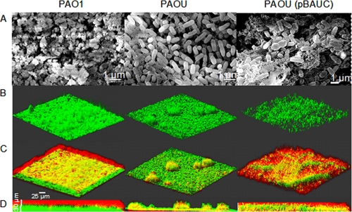
Polysaccharide matrix in P. aeruginosa biofilms. (A) SEM observations of static biofilms of the indicated strains. Magnification, ×8,500 to ×10,000. (B) 3D images resulting from CLSM observations of 48-h biofilms grown as described in the legend to Fig. 1A. The three strains contained pSMC21, and bacteria are shown in green. (C) Polysaccharides of the same biofilms as in panel B, stained with calcofluor white. This dye produced a blue light, which we artificially converted to red in our images for better contrast. Yellow results from the green (bacteria) and red (polysaccharides) overlay. (D) Side views of biofilms shown in panel C. All images are representative of five observations.

