FIG. 1.
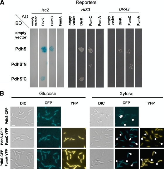
B. abortus PdhS interacts with FumC. (A) Yeast two-hybrid assay for the PdhS-FumC interaction. The fusions to either the AD or the BD of Gal4p are indicated next to the pictures. Three reporters were assayed for activation. PdhS′N and PdhS′C correspond to the N-terminal (613 aa) and C-terminal (425 aa) regions of PdhS, respectively. The PdhS-FumC interaction is positive for lacZ, HIS3, and URA3 reporters. The PdhS′N-FumC interaction is positive for HIS3 and URA3 reporters, suggesting that FumC interacts with the N-terminal part of PdhS. The divK and fumA CDS were used as positive and negative controls for the PdhS-FumC interaction, respectively. Another set of negative controls corresponded to the plasmids (“empty vector”) coding for either the Gal4p AD or the Gal4p BD, without insert. (B) Reconstruction of FumC recruitment by PdhS in C. crescentus. Three strains of C. crescentus carrying a pdhS-cfp fusion (XDB1166) under the control of the xylX promoter were constructed. In two of them, an additional fusion, either fumC-yfp (pJM080) or fumA-yfp (pJM095; negative control), is carried on a replicative plasmid. The cells were grown overnight in PYE (peptone-yeast extract) with glucose to reach a starting optical density at 600 nm (OD600) of 0.05. The medium was then supplemented with glucose (no induction) or xylose (induction) for 5 h, and the cells were subsequently immobilized on agarose pad slides and visualized by differential interference contrast (DIC) microscopy and in CFP and/or YFP channels. The fusions produced by each strain are indicated on the left. The type of observation, either DIC microscopy (Normarski) or CFP or YFP typical fluorescence, is also indicated for each culture medium. White arrows indicate the positions of some PdhS-CFP foci. All pictures are shown with the same magnification. Bar, 2 μm.

