FIG. 2.
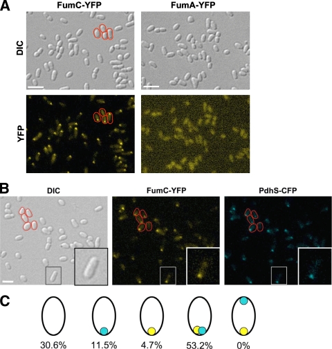
FumC-YFP is localized at the old pole in B. abortus. (A) Localization of FumC-YFP and FumA-YFP in B. abortus. The XDB1150 and XDB1153 strains, producing, respectively, FumC-YFP and FumA-YFP from fusions integrated into the B. abortus 544 chromosome, were observed during the exponential growth phase by DIC microscopy (Normarski) and for the typical fluorescence spectrum of YFP. (B) Colocalization of PdhS-CFP with FumC-YFP in B. abortus. A fumC-yfp fusion was integrated by homologous recombination at the fumC locus in the chromosome of XDB1155, a B. abortus strain in which pdhS was replaced by a pdhS-cfp allele. Observations were made by DIC microscopy (Normarski) and for typical fluorescence of YFP and CFP, as indicated. Some bacteria are surrounded with a red line in the pictures to show that the FumC-YFP and PdhS-CFP signals are polar. Bar, 2 μm. (C) Schematic drawing illustrating the proportions of cells that exhibit a localization pattern for PdhS-CFP and/or FumC-YFP. A total of 425 bacteria were examined. The polar foci of PdhS-CFP and FumC-YFP are shown in blue and yellow, respectively. It is striking that all 226 bacteria (53.2%) for which a CFP focus and a YFP focus were detectable have colocalized signals and that none present foci labeling the opposite poles. A quite large fraction of the population (30.6%) does not display any focus, whether CFP or YFP. This could be due to a low expression level of the fusions, resulting in poor sensitivity for the fluorescent signal.

