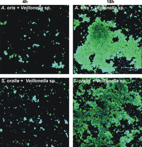
FIG. 4.
Representative confocal micrographs of 4-h (left column) and 18-h (right column) biofilms showing growth in two-species-inoculated flow cells. Top panels, Veillonella sp. plus A. oris; bottom panels, Veillonella sp. plus S. oralis. Bacterial cells were stained with species-specific fluorophore-conjugated immunoglobulin G (blue, Veillonella sp.; green, A. oris and S. oralis) and show cell-cell contact.

