FIG. 1.
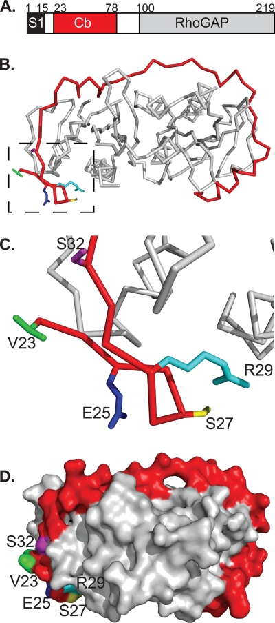
The SycE-YopE chaperone-effector complex. (A) Schematic of YopE domains. S1, signal 1; Cb, chaperone-binding region; RhoGAP, Rho GTPase activating protein domain. Residue numbers for domain boundaries are indicated. (B) Structure of the SycE-YopE(Cb) complex. The Cb region of YopE (red) and the SycE dimer (gray) are shown in C-α stick representation. Side chains that were mutated in the YopE Cb region are depicted for the following residues: V23 (green), E25 (blue), S27 (yellow), R29 (cyan), and S32 (magenta). Molecular figures were made with PyMol (http://pymol.sourceforge.net). (C) Enlarged view of boxed region in panel B. (D) Molecular surface representation of the SycE-YopE(Cb) complex. The YopE(Cb) surface is shown in red, except for surfaces formed by V23 (green), E25 (blue), S27 (yellow), R29 (cyan), and S32 (magenta). The SycE homodimer is shown in gray.

