FIG. 5.
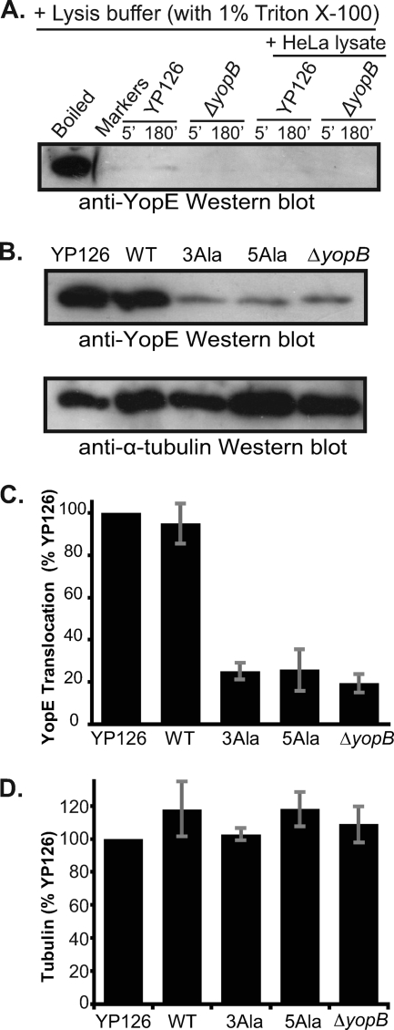
Translocation of YopE. (A) Western blot of YopE in the supernatant fraction of Y. pseudotuberculosis 126 or Y. pseudotuberculosis 126 ΔyopB incubated for 5 or 180 min in HeLa cell lysis buffer (containing 1% Triton X-100) in the absence or presence of a HeLa cell lysate. As a positive control for YopE, YP126 was incubated in PBS and lysed by boiling in SDS-PAGE sample buffer (boiled). (B) (Top) Western blot of wild-type YopE translocated into HeLa cells by Y. pseudotuberculosis 126 (YP126); wild-type YopE (WT), YopE-3Ala (3Ala), and YopE-5Ala (5Ala) translocated by Y. pseudotuberculosis 126 ΔyopE; and wild-type YopE translocated by Y. pseudotuberculosis 126 ΔyopB. Translocated fractions were separated by SDS-PAGE and probed with anti-YopE polyclonal antibodies. (Bottom) The blot at the top was stripped and reprobed with anti-α-tubulin monoclonal antibodies. (C) Quantification of YopE translocation, assessed by Western blotting. Mean values from three independent experiments carried out in triplicate are depicted. These values were normalized to the translocation level of YP126 and are expressed as percentages of the YP126 value. Gray bars report standard errors of the means. (D) Quantification of α-tubulin in translocation samples, assessed by Western blotting. Mean values from three independent experiments carried out in triplicate are depicted. These values were normalized to the level of tubulin in the YP126 sample and are expressed as percentages of the YP126 value. Gray bars report standard errors of the means.

