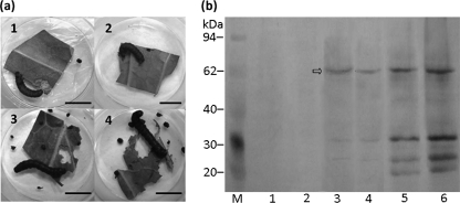
FIG. 2.
Detection of Vip3Aa1 in samples washed off the larval midgut 18 and 36 h after fourth-instar larvae of S. litura were fed with cabbage leaf discs soaked in a concentrated conidial suspension of BbV28 or BbW (negative control). (a) Larval symptoms 18 h after conidial ingestion. (1) BbV28; (2) lysate of E. coli cells expressing Vip3Aa1 (positive control); (3) BbW; (4) 0.02% Tween 80 (blank control). Bars, 1 cm. (b) Western blot of midgut samples, using a polyclonal antibody against Vip3Aa1 (arrow). Lanes 1, 3, and 5, samples washed off larval midguts after 18 h of feeding on leaf discs soaked in conidial suspensions of BbW and BbV28 and lysate of E. coli cells expressing Vip3Aa1, respectively; lanes 2, 4, and 6, same midgut samples as those in lanes 1, 3, and 5, taken after 36 h of feeding.

