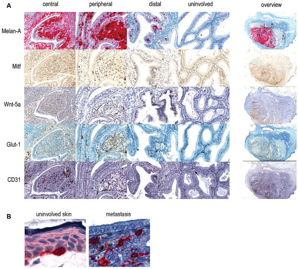
Figure 3. Immunostaining a gallbladder melanoma lesion.
(A) A gallbladder melanoma metastasis fixed in formalin and embedded in paraffin was sectioned and immunostained for Melan-A, Mitf, Wnt5A, Glut-1 and CD31. Examples of four separate regions including (i) central, (ii) peripheral, (iii) distal, and (iv) uninvolved are shown in detail, as well as an overview of the complete section, for each marker. (B) Detail of a Melan-A positive melanocyte in uninvolved skin (left) and the peripheral area of the gall bladder epithelium showing invading Melan-A positive tumor cells (right).

