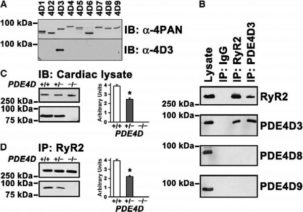
Figure 4. PDE4D3 Is a Component of the RyR2 Ca2+-Release-Channel Complex.
(A) PAN-PDE4 antibody against the conserved UCR2 domain (α-4PAN, top panel) and antibody raised against the N-terminal domain unique to PDE4D3 (α-4D3, bottom panel) were used to detect PDE4D isoforms in extracts of COS7 cells overexpressing recombinant PDE4D splice variants 1 to 9. Samples were size fractionated on 6% SDS-PAGE and blotted onto Immobilon-P membranes.
(B) Immunoblotting with splice-variant-specific anti-PDE4D3, anti-PDE4D8, and anti-PDE4D9 antibodies shows expression of all three major forms in the heart. However, immunoprecipitation of RyR2 followed by splice-variant-specific immunoblot demonstrates that only PDE4D3 is associated with RyR2. Reverse immunoprecipitation with a specific anti-PDE4D3 antibody confirms that PDE4D3 is physically associated with RyR2.
(C) Immunoblots of cardiac lysates showing amounts of RyR2 and PDE4D3 in wt, PDE4D+/−, and PDE4D−/− mice. Bar graph shows a 37% reduction of PDE4D3 in the RyR2 complex in cardiac lysates of PDE4D+/− mice relative to wt. *p < 0.05, n = 3 for each genotype. Data in (C) and (D) are mean ± SD.
(D) Coimmunoprecipitation using anti-RyR2 antibody, showing a 44% decrease of PDE4D3 bound to RyR2 in PDE4D+/− mouse hearts relative to wt. *p < 0.05, n = 3 for each genotype.

