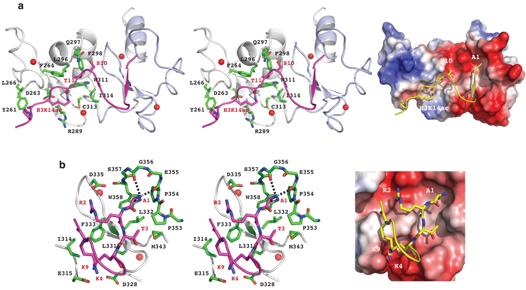
Figure 2. Structural mechanism of acetyl-lysine recognition by human DPF3b PHD12.
(a) DPF3b PHD1 recognition of H3K14ac, as illustrated in the PHD12/H3K14ac complex structure with stereo ribbon view (left) and electrostatic potential surface of the protein (right). (b) DPF3b PHD2 recognition of the N-terminal residues of the H3K14ac peptide, depicted in stereo ribbon structure (left) and protein electrostatic potential surface representation (right). The zinc atoms are highlighted as red spheres.

