
Figure 2.
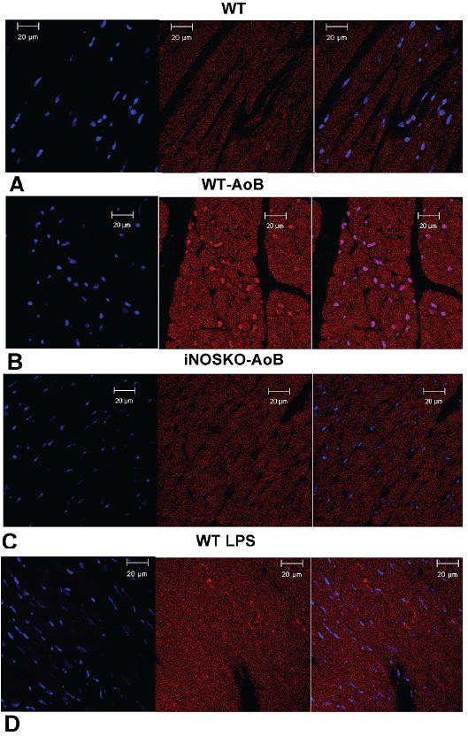
Expression of iNOS in long-term WT-AoB, iNOSKO-AoB, age-matched WT control and LPS injected WT mouse hearts detected by immunofluorescence. Representative transverse sections through A) WT heart, B) WT-AoB heart, C) iNOSKO-AoB heart and D) LPS injected WT heart. iNOS was stained using Alexa Fluor-594 (red) and nuclei were stained using DAPI (blue). The first image from the left presents the nuclei staining, the second image iNOS staining and the third is a superimposed image.