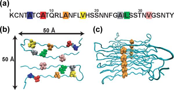
Figure 1.
hIAPP fiber model dervied from the ssNMR data. (a) The sequence of hIAPP with seven isotope labeled residues highlighted: Ala-5 (blue), Ala-8 (red), Ala-13 (orange), Val-17 (yellow), Ala-25 (silver), Leu-27 (green), and Val-32 (pink). (b) Cross section of a fiber demonstrating that the fibers are composed of two columns of random coil/β-sheet/turn/β-sheet structure. (c) Protofiber as viewed from the side with single labeled residue (Ala-13) exhibiting the formation of two isotopically labeled columns.

