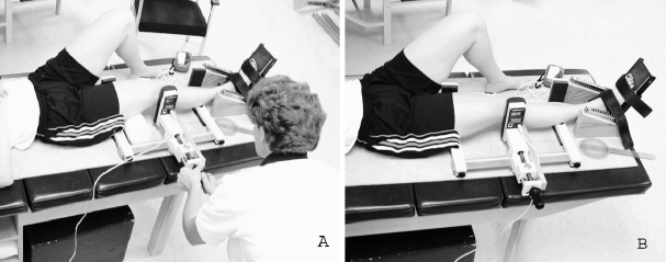
Figure 1.
Limb positioning of the tibiofemoral joint in the arthrometer. The foot plate was used to reduce femoral or tibial rotation (or both) during data collection. Force is applied to the femoral condyle, just superior to the lateral joint line. A, The medial right knee in full extension. B, The same knee is measured in 20° of flexion. A 10.16-cm foam roller was placed under the thigh to promote relaxation of the muscles at the desired angle of joint flexion when each angle was tested. A superficial electromyographic biofeedback device also ensured muscle relaxation during data collection.

