Figure 1.
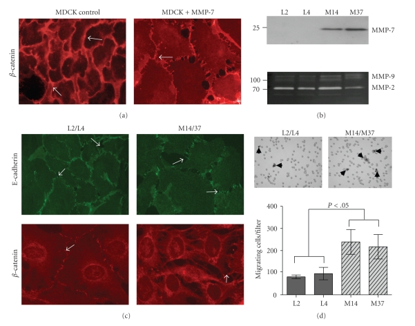
MMP-7 expression disrupts adherens junctions. (a) Representative photomicrograph (400x) of β-catenin immunofluorescent staining in MDCK cells in the absence or presence of exogenous MMP-7 (100 ng/mL). Arrows indicate junctional β-catenin. (b) Immunoblot analysis of MMP-7 (27 kDa) expression (upper panel) in empty vector control (L2 and L4) and MMP-7 (M14 and M37) transduced C57MG cell lines. Zymography (lower panel) was used to examine the expression of MMP-2 (72 kDa) and MMP-9 (92 kDa). (c) Representative photomicrographs (400x) of E-cadherin (green) and β-catenin (red) immunofluorescent staining (arrows) in the vector control (L2/L4) and MMP-7 (M14/37) expressing cells. Arrows indicate junctional E-cadherin and β-catenin. (d) The impact of MMP-7 expression on cell migration through 8 μm pore membranes (arrow heads) towards 10% serum-containing media was determined. *P < .05, as determined by ANOVA followed by Tukey's post hoc test.

