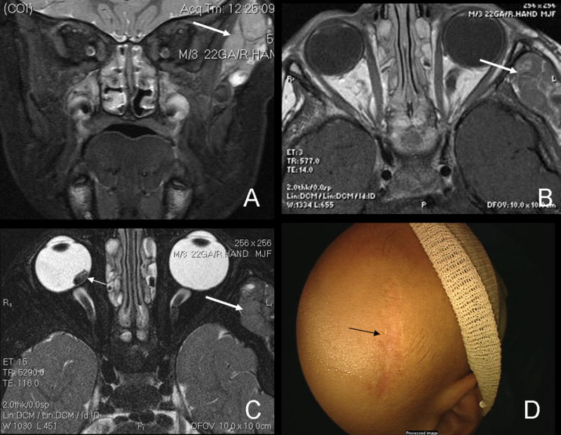
Figure 1.

Short inversion-time inversion-recovery (STIR) MRI of temporalis mass (A). The heterogeneous, well circumscribed mass which is hyperintense to muscle can also be seen on T1-weighted images (B) as well as T2 images (C), large arrows. There is no radiological evidence of extension into the globe or bones. The residual retinoblastoma tumor can be seen on the T2 image as well (small arrow, C). Post-chemotherapy for rhabdomyosarcoma, the mass has resolved, leaving only the biopsy scar (D).
