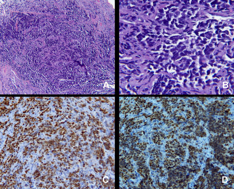
Figure 2.

Histopathology of left temporalis mass. H&E is shown demonstrating sheets of round, homogenous blue cells at 40 × magnification (A) and 400× magnification (B). The neoplastic cells are positive for rhabdomyosarcoma markers myogein (C) and myod-D1 (D), magnification 200×.
