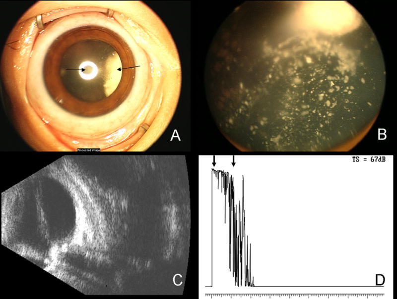
Figure 3.

Ret-Cam photograph of the patient’s right eye with advanced retinoblastoma (A). A posterior subcapsular cataract, induced by radiation, can be seen (arrow, center) as well as the white tumor around 3:00 (arrow, right). Ret-Cam photograph of the fundus of the right eye shows the advanced retinoblastoma, including inactive calcific vitreous seeds (B). B-scan ultrasound of the right eye demonstrates the shadowing generated by the calcific RB tumors (C). Diagnostic A-scan demonstrates high internal reflectivity of the calcific tumor (arrows, D).
