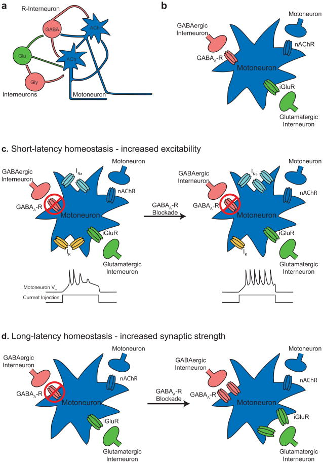
Figure 1. Homeostatic regulation of spontaneous network activity in the chick spinal cord.
When a part of the spinal cord network is blocked, activity becomes temporarily less frequent, but recovers to pre-block levels. Here we provide schematics of the changes that take place after activity blockade.
a. Schematic of the circuits that mediate activity in the developing spinal cord. Neurons are color-coded by the transmitter they release. ACh, acetylcholine, blue; Glu, glutamate, green; Gly, glycine, pink; GABA, pink.
b. Motor neurons provide a crucial drive in the generation of activity. They receive input from other motor neurons and from interneurons. nAChR, nicotinic acetylcholine receptor; GABAA-R, GABAA receptor; iGluR, ionotropic glutamate receptor.
c. When GABAA receptors are blocked in ovo (left), activity becomes temporarily less frequent but recovers 11. After 12 hours of GABAA-R blockade, motor neurons become more excitable, an effect that is mediated by an increase in the density of sodium current and a decrease in the density of potassium current111 (right). Bottom: schematics illustrating an increase in motor neuron excitability, with the bottom curve showing current injection into a motor neuron, and the top curve showing membrane potential. A more excitable motor neuron fires more action potentials in response to the same stimulus (right). INa, sodium current, represented by sodium channels; IK, potassium current, represented by potassium channels.
d. When GABAA receptors are blocked for long periods (24–48 hours) glutamatergic and GABAergic postsynaptic currents in motor neurons increase in size110. The exact mechanisms underlying this increase in postsynaptic current are not fully understood, but are schematized here as increases in the number of glutamate and GABAA receptors.

